Definition
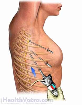
Thoracic surgery is done on the chest, but it does not involve surgery on the heart. With robot-assisted thoracic procedures, the doctor guides small robotic arms through keyhole incisions.

Reasons for Procedure
Robot-assisted thoracic procedures are considered for surgeries that:
- Require precision
- Do not require open access
Some thoracic surgeries that have been successfully performed using robotic techniques include:
- Thymectomy —removal of the thymus gland
- Lobectomy —removal of a lung lobe
- Esophagectomy —removal of the esophagus
- Mediastinal tumor resection —removal of tumors located in the mediastinum (the part of the chest cavity that separates the lungs)
- Sympathectomy—cauterizing a portion of the sympathetic nerve
Compared to more traditional procedures, robotic-assisted surgery may result in:
- Less scarring
- Reduced recovery times
- Less risk of infection
- Less blood loss
- Reduced trauma to the body
- Shorter hospital stay
- Faster recovery
Possible Complications
Complications are rare, but no procedure is completely free of risk. If you are planning to have a robot-assisted thoracic procedure, your doctor will review a list of possible complications, which may include:
- Bleeding
- Infection
- Collection of air or gases in the lung cavity ( pneumothorax)
- Collapsed lung
- Need for a prolonged artificial respiration on a ventilator (breathing machine)
- Damage to neighboring organs or structures
- The need to switch to traditional surgical methods (eg, traditional laparoscopic or open surgery)
- Anesthesia-related problems
- Nerve damage
Some factors that may increase the risk of complications include:
- Advanced age
- Obesity
- Smoking
- Diabetes
- Excessive alcohol intake
- Use of certain medicines
Be sure to discuss these risks with your doctor before the procedure.
What to Expect
Prior to Procedure
Depending on the reason for your surgery, your doctor may do the following:
- Physical exam
- Blood tests and urine tests
- Chest x-ray —a test that uses radiation to take a picture of structures inside the chest
- Pulmonary function test —a test to assess lung function
- Upper GI series —x-ray of the esophagus, stomach, and part of the small intestines after swallowing a barium solution
- Electrocardiogram (ECG, EKG) —a test that records the electrical currents passing through the heart muscle
- Ultrasound —a test that uses sound waves to visualize the inside of the chest
- CT scan —a type of x-ray that uses a computer to create images of structures inside the chest
- MRI scan —a test that uses powerful magnets and radiowaves to create images of structures inside the chest
- Upper endoscopy —a lighted tube equipped with a camera is used to visualize the inside of the esophagus, stomach, and part of the small intestines
Leading up to the procedure:
- Talk to your doctor about your medicines. You may be asked to stop taking some medicines up to one week before the procedure, like:
- Anti-inflammatory drugs (eg, aspirin )
- Blood thinners such as clopidogrel (Plavix) or warfarin (Coumadin)
- Take antibiotics if instructed.
- Follow a special diet if instructed.
- Take a laxative and/or use an enema to clean out your intestines if instructed.
- Shower the night before using antibacterial soap if instructed.
- Arrange for someone to drive you home from the hospital. Also, have someone to help you at home.
- Eat a light meal the night before. Do not eat or drink anything after midnight.
Anesthesia
General anesthesia will be used. It will block any pain and keep you asleep through the surgery.
Description of the Procedure
You will be connected to a ventilator. This is a machine that moves air in and out of your lungs. Next, the doctor will cut several keyhole openings in the chest wall between the ribs. One or more chest tubes may be placed into the side of the chest. These tubes will be used to drain fluid and monitor air leakage. A needle may be used to inject carbon dioxide gas into the chest cavity. The gas will make it easier for the doctor to see internal structures.
The doctor will then pass a small camera, called an endoscope, through one of the incisions. The camera will light, magnify, and project the structures onto a video screen. The camera will be attached to one of the robotic arms. The other arms will hold instruments for grasping, cutting, dissecting, and suturing. These may include:
- Forceps
- Scissors
- Dissectors
- Scalpels
While sitting at a console near the operating table, the doctor will look through lenses at magnified 3D images of the inside of the body. Another doctor will stay by the table to adjust the camera and tools. With joystick-like controls and foot pedals, the doctor will guide the robotic arms and tools to remove organs and tissue. After the tools are removed, the doctor will use sutures or staples to close the surgical area.
Immediately After Procedure
If you are doing well, the breathing tube will be removed. Later, the chest tubes will be removed.
How Long Will It Take?
About 1-4 hours (depending on the procedure)
How Much Will It Hurt?
You will have pain during recovery. Your doctor will give you pain medicine. You may also feel discomfort from the gas used during the procedure. This can last up to three days.
Average Hospital Stay
This procedure is done in a hospital setting. The usual length of stay is a few days. Your doctor may choose to keep you longer if you have any problems.
Post-procedure Care
At the Hospital
While you are recovering at the hospital, you may receive the following care:
- Assistance sitting up and moving around soon after surgery
- Instructions on what you should eat and how to restrict your activity
- Nutrition through a feeding tube in the days after surgery (You will gradually progress from a liquid to a solid diet.)
- Directions on how to do deep breathing and coughing exercises
At Home
When you return home, do the following to help ensure a smooth recovery:
- Take antibiotics to prevent infection if instructed.
- Avoid certain medicines.
- Resume normal activities (eg, daily walks) soon. This will promote healing.
- Wash the incisions with mild soap and water.
- Ask your doctor about when it is safe to shower, bathe, or soak in water.
- Limit certain activities (eg, driving, working, doing strenuous exercise) until you have recovered.
- Be sure to follow your doctor’s instructions.
Depending on the procedure, you should recover within a few weeks.
Call Your Doctor
After you leave the hospital, contact your doctor if any of the following occurs:
- Cough or shortness of breath
- Coughing up yellow, green, or bloody mucus
- New chest pain
- Signs of infection, including fever and chills
- Redness, swelling, increasing pain, excessive bleeding, or discharge from an incision site
- Difficulty urinating, such as pain, burning, urgency, frequency, or bleeding
- Pain and/or swelling in your feet, calves, or legs
- Persistent nausea, vomiting, and/or diarrhea
- Headache, feeling faint or dizzy
- Other worrisome symptoms
In case of an emergency, call for medical help right away.
