সংজ্ঞা
An echocardiogram uses sound waves called ultrasound to look at the size, shape, and motion of the heart.
The test shows:
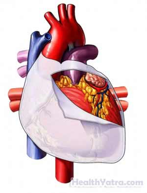
- Four chambers of the heart
- Heart valves and the walls of the heart
- Blood vessels entering and leaving the heart
- The sac that surrounds the heart

In addition to this standard test, there are specialized echocardiograms:
- Contrast echocardiogram—A solution is injected into the vein and can be seen in the heart.
- Stress echocardiogram—This records the heart’s activity during a cardiac stress test.
- Echocardiogram with Doppler ultrasound—This helps your doctor assess blood flow.
- Transesophageal echocardiogram—To provide clear images of the heart, the ultrasound device is put down your throat. Your doctor may need to use this test depending on what part of the heart needs to be viewed. If you have the following conditions, you may need this test, rather than the standard echocardiogram:
- Certain lung diseases
- স্থূলতা
পরীক্ষার কারণ
An echocardiogram may be used to:
- Evaluate a heart murmur
- Diagnose valve conditions
- Find changes in the heart’s structure
- Assess motion of the chamber walls and damage to the heart muscle after a heart attack
- Assess how different parts of the heart work in people with chronic heart disease
- Determine if fluid is collecting around the heart
- Identify growths in the heart
- Assess and monitor birth defects
- Test blood flow through the heart
- Assess heart or major blood vessel damage caused by trauma
- Test heart function and diagnose heart and lung problems in very ill patients
- Assess chest pain
- Look for blood clots within heart chambers
সম্ভাব্য জটিলতা
এই পরীক্ষার সাথে যুক্ত কোন বড় জটিলতা নেই।
কি আশা করা যায়
পরীক্ষার আগে
আপনার ডাক্তার নিম্নলিখিত করতে পারেন:
- শারীরিক পরীক্ষা
- Electrocardiogram (ECG, EKG)—a test that records the heart’s activity by measuring electrical currents through the heart muscle
পরীক্ষার বর্ণনা
A gel is put on your chest. This gel helps the sound waves travel. A small, hand-held device called a transducer is pressed against your skin. The transducer sends sound waves toward your heart. The sound waves are then reflected back to the device. The waves are converted into electrical impulses. These impulses become an image on the screen.
Still images or videotape moving images can be captured. To get clearer and more complete images, the transducer may be moved to different areas of your chest. You may be asked to change positions and slowly inhale, exhale, or hold your breath.
পরীক্ষার পর
The gel is wiped from your chest.
কতক্ষণ এটা লাগবে?
30-60 minutes
এটা আঘাত করবে?
না
ফলাফল
The images are analyzed. Based on the findings, your doctor may recommend treatment or further testing.
আপনার ডাক্তারকে কল করুন
After the test, call your doctor if you have worsening heart-related symptoms.
